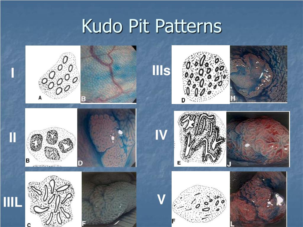
Kudo Pit Pattern
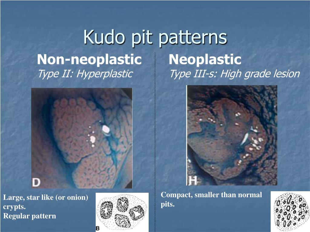
Kudo Pit Pattern - Web kudo's pit pattern classification is an accurate diagnostic method for the differentiation of neoplastic colorectal lesions. Web the kudo pit pattern with narrow band imaging (nbi) has high sensitivity and specificity for distinguishing adenomas (type iii/iv) from hyperplastic polyps (type i/ii), particularly when used by experts. Kudo et al[5] classified colorectal polyps according to their appearance, structure and staining patterns. Called the surface microstructure “pit pattern”, and they came to find a constant rule since “pit pattern” was analyzed corresponding to the pathological features and diagnosis. Web a value of 4 or higher in the kudo classification turned out to be the best parameter to differentiate malignant lesions from benign ones for lesions larger than 1 cm and laterally spreading tumors, with a sensitivity of 91.2% and a specificity of 70.4%. 6 type i describes small round pits that typically represent normal histology. The kudo pit pattern highlighted by kudo and colleagues22 describes “pit patterns” to identify neoplastic and nonneoplastic polyps with the use of magnifying endoscopy with (chromoendoscopy) or without (nbi) dye spray. Web for microvascular pattern evaluation, the nbi international colorectal endoscopic (nice) classification and the japanese nbi expert team (jnet) classification are the most widely disseminated, while the kudo classification is the gold standard for pit pattern assessment using crystal violet [15,24]. Publication bias is significant in the current available literature. Web pit pattern in colorectal neoplasia: Web the kudo pit pattern (figure 5), with the use of dye chromoendoscopy, has been found to be highly accurate for predicting superficial (type vi) and deep submucosal invasion (type vn). Publication bias is significant in the current available literature. Web kudo's pit pattern classification is an accurate diagnostic method for the differentiation of neoplastic colorectal lesions. 6 type i describes small round pits that typically represent normal histology. Type ⅰ pits appear as roundish pits; Web the kudo pit pattern classification system uses magnification colonoscopy with dye spray (chromoendoscopy) to categorize polyps based on the appearance of their pits, which are crypt openings. This scheme broadly categorizes pit patterns into 7 types based on the pit appearance and structure (figure 3 ). Of surgery and gastroenterology, akita red cross hospital, japan. S kudo 1 , c a rubio , c r teixeira , h kashida , e kogure. Called the surface microstructure “pit pattern”, and they came to find a constant rule since “pit pattern” was analyzed corresponding to the pathological features and diagnosis. Web kudo pit pattern classification. Web the kudo pit pattern can be assessed on colonoscopy and predicts the presence or absence of neoplasia and malignancy. Kudo et al[5] classified colorectal polyps according to their appearance, structure and staining patterns. The kudo pit pattern highlighted by kudo and colleagues22 describes “pit patterns” to identify neoplastic and nonneoplastic polyps with the use. Web kudo's pit pattern classification is an accurate diagnostic method for the differentiation of neoplastic colorectal lesions. Web we recommend that the surface characteristics of a polyp should be described using a classification system such as the nice nbi or kudo pit pattern classification. The kudo pit pattern highlighted by kudo and colleagues22 describes “pit patterns” to identify neoplastic and. Web we recommend that the surface characteristics of a polyp should be described using a classification system such as the nice nbi or kudo pit pattern classification. Kudo's pit pattern classification is an accurate diagnostic method for the differentiation of neoplastic colorectal lesions. Web a value of 4 or higher in the kudo classification turned out to be the best. Web the kudo pit pattern can be assessed on colonoscopy and predicts the presence or absence of neoplasia and malignancy. Type ⅰ pits appear as roundish pits; Web we recommend that the surface characteristics of a polyp should be described using a classification system such as the nice nbi or kudo pit pattern classification. 12 however, assessing the kudo pit. Web the kudo pit pattern with narrow band imaging (nbi) has high sensitivity and specificity for distinguishing adenomas (type iii/iv) from hyperplastic polyps (type i/ii), particularly when used by experts. Called the surface microstructure “pit pattern”, and they came to find a constant rule since “pit pattern” was analyzed corresponding to the pathological features and diagnosis. Web evaluating 2693 lesions,. Publication bias is significant in the current available literature. The opening to each crypt in the epithelium is known as a pit. Web kudo's pit pattern classification is an accurate diagnostic method for the differentiation of neoplastic colorectal lesions. Kudo et al[5] classified colorectal polyps according to their appearance, structure and staining patterns. Kudo's pit pattern classification is an accurate. Web a magnifying endoscope was developed with the aim of applying the findings obtained in these studies in vivo. Web kudo's pit pattern classification is an accurate diagnostic method for the differentiation of neoplastic colorectal lesions. 6 type i describes small round pits that typically represent normal histology. Publication bias is significant in the current available literature. This scheme broadly. Web kudo's pit pattern classification was composed seven type pit pattern. Kudo and colleagues first highlighted the feasibility of examining and classifying pit patterns to distinguish type of polyps by using magnifying endoscopy. Publication bias is significant in the current available literature. Called the surface microstructure “pit pattern”, and they came to find a constant rule since “pit pattern” was. Stellar or papillary pits are type ii, and roundish tubular pits are classified as type iii (smaller than. Kudo's pit pattern classification is an accurate diagnostic method for the differentiation of neoplastic colorectal lesions. Web pit pattern in colorectal neoplasia: Web evaluating 2693 lesions, kudos pit pattern v was the strongest factor associated with overt submucosal invasive cancer (odds ratio. Web a magnifying endoscope was developed with the aim of applying the findings obtained in these studies in vivo. Publication bias is significant in the current available literature. Web we recommend that the surface characteristics of a polyp should be described using a classification system such as the nice nbi or kudo pit pattern classification. Web kudo's pit pattern classification. Web kudo pit pattern classification. Web kudo's pit pattern classification is an accurate diagnostic method for the differentiation of neoplastic colorectal lesions. Web evaluating 2693 lesions, kudos pit pattern v was the strongest factor associated with overt submucosal invasive cancer (odds ratio [or], 1.42; Stellar or papillary pits are type ii, and roundish tubular pits are classified as type iii (smaller than. 6 type i describes small round pits that typically represent normal histology. Of surgery and gastroenterology, akita red cross hospital, japan. Web for microvascular pattern evaluation, the nbi international colorectal endoscopic (nice) classification and the japanese nbi expert team (jnet) classification are the most widely disseminated, while the kudo classification is the gold standard for pit pattern assessment using crystal violet [15,24]. Web the kudo pit pattern classification system uses magnification colonoscopy with dye spray (chromoendoscopy) to categorize polyps based on the appearance of their pits, which are crypt openings. Web the kudo pit pattern with narrow band imaging (nbi) has high sensitivity and specificity for distinguishing adenomas (type iii/iv) from hyperplastic polyps (type i/ii), particularly when used by experts. Web the kudo pit pattern (figure 5), with the use of dye chromoendoscopy, has been found to be highly accurate for predicting superficial (type vi) and deep submucosal invasion (type vn). Web kudo’s pit pattern classification states morphological pit patterns of the lieberkühn crypts by analyzing the superficial mucosa, predicting the histology of colorectal lesions. Kudo and colleagues first highlighted the feasibility of examining and classifying pit patterns to distinguish type of polyps by using magnifying endoscopy. Called the surface microstructure “pit pattern”, and they came to find a constant rule since “pit pattern” was analyzed corresponding to the pathological features and diagnosis. Web kudo describes five different ‘pit patterns’. Web a magnifying endoscope was developed with the aim of applying the findings obtained in these studies in vivo. The kudo pit pattern highlighted by kudo and colleagues22 describes “pit patterns” to identify neoplastic and nonneoplastic polyps with the use of magnifying endoscopy with (chromoendoscopy) or without (nbi) dye spray.
Endoscopic Recognition and Resection of Malignant Colorectal Polyps

Pit pattern classification of colorectal neoplasia (Adapted from Tanaka

Kudo classification of pit patterns with photographic correlation of

Kudo classification of pit patterns with photographic correlation of

Kudo's pit pattern classification was composed seven type pit pattern

Figure 1 from Quantitative analysis and development of a computeraided
PPT EQUIP Training session 2 PowerPoint Presentation, free download

Kudo's pit pattern classification was composed seven type pit pattern
PPT EQUIP Training session 2 PowerPoint Presentation, free download

Classificazione del pit pattern secondo Kudo e raffronto tra visione
Web Kudo's Pit Pattern Classification Was Composed Seven Type Pit Pattern.
Web Kudo's Pit Pattern Classification Is An Accurate Diagnostic Method For The Differentiation Of Neoplastic Colorectal Lesions.
Web Pain Visual Analog Scale (Vas) And Emotional Scale Were Used To Evaluate Pain And Emotions In Both Groups Of Patients.
15 The Different Pit Patterns Include Round/Normal (Type I), Asteroid (Type Ii), Tubular Or Round Pit That Is Smaller Than The Normal (Type Iiis.
Related Post: