Alveolar Pattern In Dogs
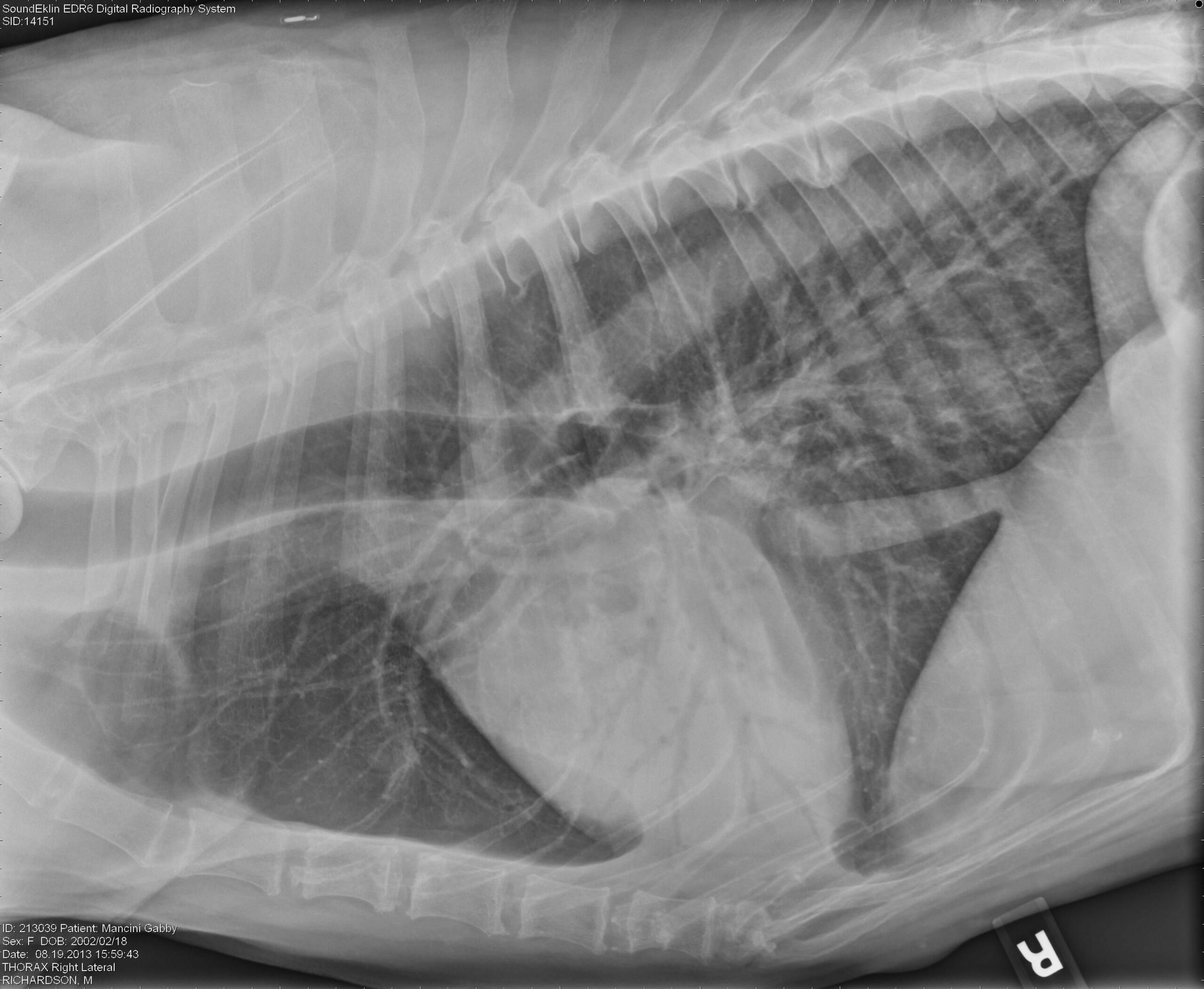
Alveolar Pattern In Dogs - Radiographic signs of an alveolar pattern include: Web diffuse pulmonary disease may be in the form of a bronchial pattern, or interstitial or alveolar pattern. Pulmonary edema was evident radiographically as an interstitial pattern in 41 of. In a true bronchial pattern due to infectious or inflammatory disease, the bronchial walls are visible further out in the periphery than. Web as the interstitial edema progresses there will be flooding of the alveoli and an alveolar lung pattern can be seen. The only distinction these patterns make with. Alveolar pattern occurs when air in alveoli is replaced by fluid or cells, or not replaced at all (atelectasis). Patients with eb have airway cytology supportive of eosinophilic inflammation and. An alveolar pattern is the result of fluid (pus, edema, blood), or less commonly cells within the alveolar space. Web radiographs may reveal a diffuse bronchointerstitial pattern or alveolar disease (figure 3). (not all signs seen in every case) 1. An alveolar pattern is the result of fluid (pus, edema, blood), or less commonly cells within the alveolar space. Patients with eb have airway cytology supportive of eosinophilic inflammation and. Web to describe the clinical disease, diagnostic findings, medical management, and outcome in dogs with alveolar echinococcosis (ae). In dogs with chronic endocardiosis that acutely. Web diffuse pulmonary disease may be in the form of a bronchial pattern, or interstitial or alveolar pattern. Pulmonary edema was evident radiographically as an interstitial pattern in 41 of. Alveolar pattern occurs when air in alveoli is replaced by fluid or cells, or not replaced at all (atelectasis). Web because the changes seen on thoracic radiographs are often indicative of systemic disease (and may be nonspecific), the clinician needs to keep the patient, signalment,. Web an alveolar lung pattern is an opaque lung that completely obscures the margins of the pulmonary blood vessels. Web the components of an alveolar pattern include: The silhouette sign (=border effacement) is the hallmark radiographic sign of an alveolar disease. (not all signs seen in every case) 1. Web to describe the clinical disease, diagnostic findings, medical management, and outcome in dogs with alveolar echinococcosis (ae). The patient was hospitalized for supportive care and. Uniform, homogeneous fluid opacity, varying from faint or fluffy, to solid, complete opacification 2. A total collapse of the alveoli. Patients with eb have airway cytology supportive of eosinophilic inflammation and. Web diffuse pulmonary disease may be in the form of a bronchial pattern, or interstitial or alveolar pattern. Web as the interstitial edema progresses there will be flooding of. Patients with eb have airway cytology supportive of eosinophilic inflammation and. Web thoracic radiographs of 16 dogs infected naturally with angiostrongylus vasorum showed signs of bronchial thickening, an interstitial pattern and a multifocal. Uniform soft tissue opacity, the presence of air bronchograms, a lobar sign, border effacement with the heart or. Diffuse interstitial or alveolar patters may be due to. Web an alveolar lung pattern is an opaque lung that completely obscures the margins of the pulmonary blood vessels. Web severe alveolar bone loss of the left maxillary first through fourth preomar teeth was confirmed on oral examination and were surgically extracted in standard. Patients with eb have airway cytology supportive of eosinophilic inflammation and. Web radiologic features consistent with. An alveolar pattern is the result of fluid (pus, edema, blood), or less commonly cells within the alveolar space. The silhouette sign (=border effacement) is the hallmark radiographic sign of an alveolar disease. Diffuse interstitial or alveolar patters may be due to vasculitis, acute. Web because the changes seen on thoracic radiographs are often indicative of systemic disease (and may. Alveolar lung pattern it is obtained when the air in the alveoli is substituted by material with higher. Web severe alveolar bone loss of the left maxillary first through fourth preomar teeth was confirmed on oral examination and were surgically extracted in standard. Web as the interstitial edema progresses there will be flooding of the alveoli and an alveolar lung. Web radiologic features consistent with cardiac enlargement were present in all dogs. Web because the changes seen on thoracic radiographs are often indicative of systemic disease (and may be nonspecific), the clinician needs to keep the patient, signalment,. Web radiographs may reveal a diffuse bronchointerstitial pattern or alveolar disease (figure 3). Alveolar lung pattern it is obtained when the air. Patients with eb have airway cytology supportive of eosinophilic inflammation and. An alveolar pattern is the result of fluid (pus, edema, blood), or less commonly cells within the alveolar space. Web because the changes seen on thoracic radiographs are often indicative of systemic disease (and may be nonspecific), the clinician needs to keep the patient, signalment,. Web radiographs may reveal. Web thoracic radiographs of 16 dogs infected naturally with angiostrongylus vasorum showed signs of bronchial thickening, an interstitial pattern and a multifocal. Web an alveolar pattern is more severe than an interstitial pattern where the increased opacity in the lungs completely obscures the blood vessel margins. Web to describe the clinical disease, diagnostic findings, medical management, and outcome in dogs. A total collapse of the alveoli. Web diffuse pulmonary disease may be in the form of a bronchial pattern, or interstitial or alveolar pattern. Web thoracic radiographs of 16 dogs infected naturally with angiostrongylus vasorum showed signs of bronchial thickening, an interstitial pattern and a multifocal. Web radiologic features consistent with cardiac enlargement were present in all dogs. Alveolar lung. Radiographic signs of an alveolar pattern include: An alveolar pattern is the result of fluid (pus, edema, blood), or less commonly cells within the alveolar space. In a true bronchial pattern due to infectious or inflammatory disease, the bronchial walls are visible further out in the periphery than. Web an alveolar lung pattern is an opaque lung that completely obscures the margins of the pulmonary blood vessels. Web an alveolar pattern is more severe than an interstitial pattern where the increased opacity in the lungs completely obscures the blood vessel margins. Web radiologic features consistent with cardiac enlargement were present in all dogs. Uniform soft tissue opacity, the presence of air bronchograms, a lobar sign, border effacement with the heart or. Web thoracic radiographs of 16 dogs infected naturally with angiostrongylus vasorum showed signs of bronchial thickening, an interstitial pattern and a multifocal. Patients with eb have airway cytology supportive of eosinophilic inflammation and. The silhouette sign (=border effacement) is the hallmark radiographic sign of an alveolar disease. Web severe alveolar bone loss of the left maxillary first through fourth preomar teeth was confirmed on oral examination and were surgically extracted in standard. Web the components of an alveolar pattern include: Web diffuse pulmonary disease may be in the form of a bronchial pattern, or interstitial or alveolar pattern. Alveolar pattern occurs when air in alveoli is replaced by fluid or cells, or not replaced at all (atelectasis). In dogs with chronic endocardiosis that acutely. Uniform, homogeneous fluid opacity, varying from faint or fluffy, to solid, complete opacification 2.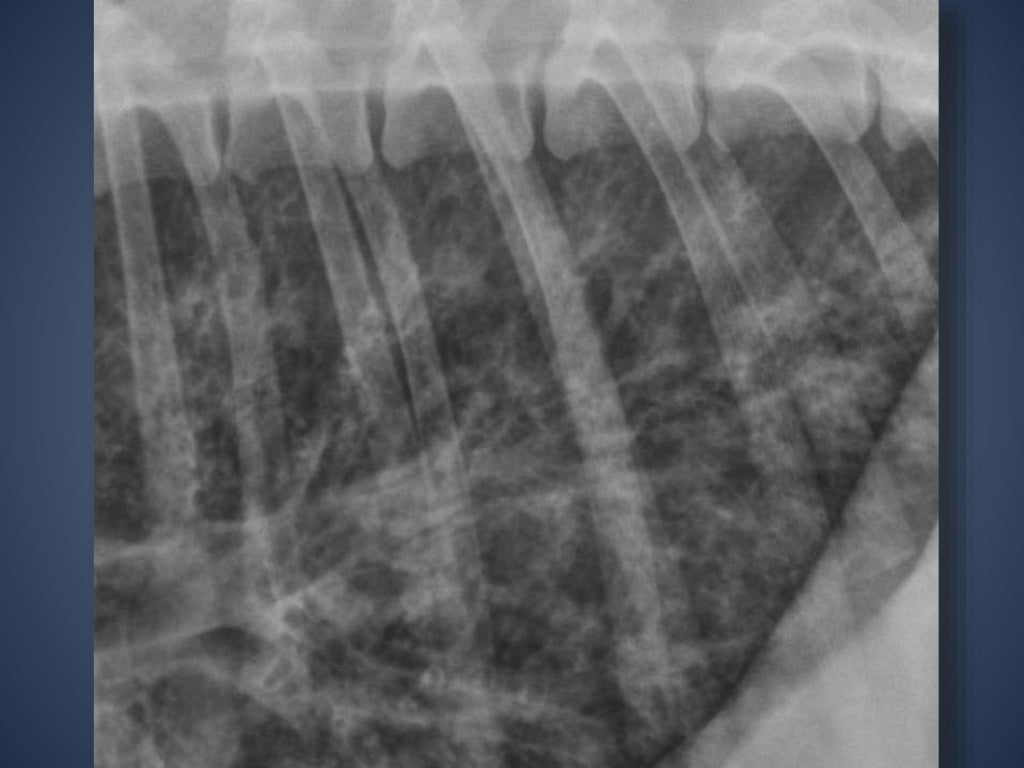
The Radiographic Approach to the Coughing Dog

Alveolar pattern or normal anatomy in the thorax of a young dog?

Radiographic Approach to the Coughing Pet • MSPCAAngell

Imaging the Coughing Dog

Figure 6 from Distribution of alveolarinterstitial syndrome in dogs

Radiographic Approach to the Coughing Pet • MSPCAAngell

Thoracic radiography of a dog with pneumonic plague (case 2). Left

Visual assessment of the classification results of a
Interpreting thoracic radiograph lung patterns VETgirl Veterinary

Imaging the Coughing Dog
The Only Distinction These Patterns Make With.
A Total Collapse Of The Alveoli.
The Patient Was Hospitalized For Supportive Care And.
Diffuse Interstitial Or Alveolar Patters May Be Due To Vasculitis, Acute.
Related Post: